
RESEARCH
Epithelial cell-cell junction mechanics
Adult epithelial cells often experience strains of tens to a few hundred percent at a wide spectrum of strain rates. They have many mechanisms to dissipate internal stress to avoid damage, often via cytoskeleton remodeling and cell-cell adhesion enhancement. These mechanisms differ in time scale and play different roles in either dissipating the strain-induced stresses or strengthening the internal cellular structures. Our lab aim to study the strain rate-dependent mechanical behavior of the cell-cell adhesion, particularly we hope to determine what coping mechanisms are at play across the spectrum of strain rates, and about how stress relaxation by the cytoskeleton coordinates with adhesion enhancement in the events leading up to rupture. This knowledge is critical in understanding various disease pathology and therapeutics.
We study this process at the single cell-cell interface. Our group developed a single-cell adhesion micro tensile tester platform. Two movable islands, supported with beams of known stiffness, are mechanically coupled through the formation of a mature junction between epithelial cells on each island. Integrating the microstructure with an atomic force microscopy enables us to stretch the cell pair with controlled strain rates, while the deformation of the supporting beams informs the resultant stress at the cell-cell junction.
To the best of our knowledge, this is the first platform that allows in situ investigation of stress-strain characteristics of a mature cell-cell junction through defined strains and strain rates. With SCAµTT, we reveal some interesting biophysical phenomena at the single cell-cell junction that were previously not possible to observe using existing techniques. We showed that cytoskeleton growth can effectively relax intercellular stress between an adherent cell pair in a strain-rate dependent manner. Along with cadherin clustering-induced bond strengthening, it prevents failure to occur at low strain rates. At high strain rates, insufficient relaxation leads to stress accumulation which results in cell-cell junction rupture. Collectively, our characterization of the strain rate-dependent mechanical behavior of the cell-cell junction builds the foundation for a new mechanistic understanding of junction adaptation to external load, and potentially the spatiotemporal coordination of participating molecules at the cell-cell junction.
Role of desmosome cell-cell junctions in cell mechanics
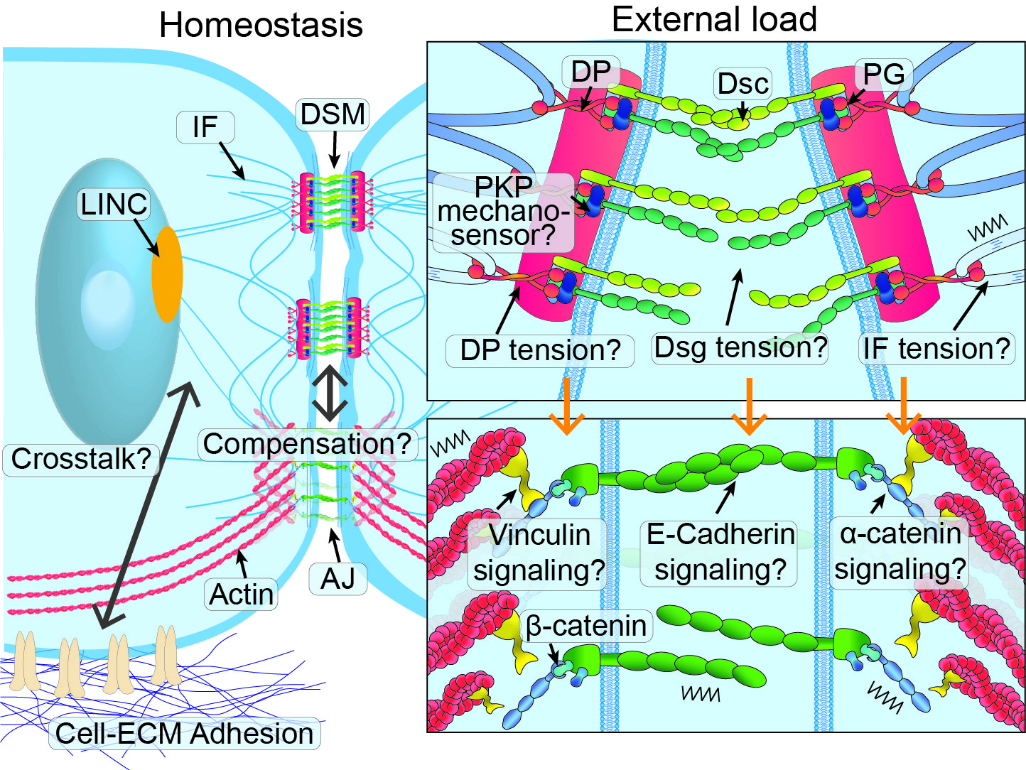
We aim to understand mechanotransduction of cell-cell adhesions under tensioned environments. My lab’s current focus is on the less studied desmosome (DSM) adhesions in epithelial cells. DSMs resist mechanical stress and are regarded as an essential signal regulator by responding to mechanical cues in development and in many diseases of the skin and heart. Understanding its transduction mechanism is of great importance to human health. While significant progress has been made in identifying potential load-bearing elements within DSM, much remains unknown about the roles DSM and its associated components play in epithelial mechanics and mechanotransduction. To address these gaps, we will focus on two major research questions: 1) investigating the role DSMs play in maintaining the mechanical integrity of cell-cell junctions and 2) determining its potential in transducing mechanical cues at the junction in coordination with molecules in adherens junctions (AJs) and the actin network.
Disruption of cell-cell junctions in disease
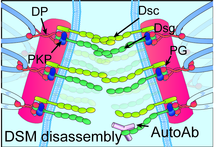
Pemphigus vulgaris (PV) is a potentially life-threatening autoimmune blistering disease that affects the skin and mucous membranes and serves as an excellent model for the study of human organ-specific autoimmunity. PV is characterized by the presence of autoantibodies (autoAbs) directed against keratinocyte surface antigens, primarily the desmosomal cadherins desmoglein 3 (Dsg3) and Dsg1. Binding of these autoAbs leads to desmosome internalization, keratin retraction, cell dissociation, and ultimately blister formation. The bulk of research in PV to date has focused on immune mechanisms upstream of autoAb binding. Consequently, therapeutic interventions in PV have mostly relied on non-specific immunosuppression, which is often accompanied by detrimental long-term side effects. However, the biophysical aspects of the cell behavior after autoAb binding are only recently being explored thanks to the advancement of innovative technologies applied to fundamental biological questions at the cellular level. We aim to study the biophysical process of cell-cell adhesion dissociation by examining the mechanics of the cell-cell adhesion at the cellular level. We expect to delineate how antibody induces the disassembly of a desmosome and how subsequent signaling pulls neighboring cells apart either via cell contractility or from external deformation.
